 BlitZ
BlitZ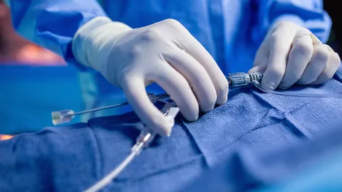
Medical imaging allows physicians to view structures inside the body without surgery. X-rays, ultrasound, computed tomography (CT), and magnetic resonance imaging (MRI) each produce these images, and they help care teams study bones, organs, and blood vessels. Interventional radiology builds on these tools, pairing real-time imaging with small instruments that physicians guide through the body.
What Is Interventional Radiology?
Interventional radiology is a type of medical specialty, and it is one in which physicians use imaging technology to guide certain procedures. Rather than making large incisions, an interventional radiologist may insert a thin needle, wire, or flexible tube called a catheter. Live images from fluoroscopy, ultrasound, CT, or MRI show where the instrument is located as it moves toward the targeted area.
The specialty differs from diagnostic radiology, which focuses primarily on capturing and interpreting images. Interventional radiologists also interpret images, but they can use that information to carry out procedures during the same session. The imaging allows the physician to follow blood vessels or other pathways to a specific location.
What Does It Detect?
Interventional radiology can play a role in both identifying and addressing health concerns. Image-guided techniques may allow physicians to examine areas that are difficult to evaluate with standard scans alone. There are some common areas that interventional radiologists can assess, including:
- Blood Vessels: Narrowing, blockages, bulges known as aneurysms, or abnormal connections between arteries and veins
- Organs: Masses or growths in the liver, kidneys, lungs, thyroid, or other tissues
- Bleeding Sources: Internal bleeding that may be located with specialized contrast imaging
- Fluid collections: Abscesses or buildups of fluid in the abdomen, chest, or other areas
A physician may also request an image-guided biopsy, and this involves removing a small tissue sample for laboratory analysis. The results can give the care team information, helping shape the next steps in a treatment plan. A diagnosis is made by the patient’s physicians after reviewing imaging, lab results, and medical history together.
What Approaches Fall Under It?
The specialty includes a broad range of procedures. The specific approach depends on the condition being evaluated and the physician’s assessment. Angiography uses contrast dye and X-ray imaging to display blood vessels. When narrowing is present, a physician may perform angioplasty, which involves inflating a small balloon inside the vessel, sometimes followed by placing a mesh tube called a stent. Embolization is a technique that intentionally blocks blood flow to a specific area, such as a bleeding vessel or certain growths.
Endovenous ablation uses heat from laser or radiofrequency energy to close veins that are not functioning as expected. Sclerotherapy involves injecting a solution into smaller veins to close them. Interventional radiologists may place drainage catheters to remove fluid from the body. They also insert ports and central lines, which provide long-term access to veins for medications or other therapies. Some techniques deliver treatment directly to a growth.
Seek Professional Radiology Care
Interventional radiology procedures may begin with a referral from another physician, followed by a review of prior imaging and medical history. Before a procedure, the care team can request blood work, provide instructions about eating and drinking, and review current medications, particularly blood thinners. Follow-up imaging or appointments may help the care team monitor progress, and they help determine whether additional steps are appropriate.